What is an MRI scan?
MRI is an abbreviation for magnetic resonance imaging. This type of scan uses magnetic fields and radio waves in a non-invasive way to diagnose problems inside the body. It produces detailed images of organs, bones and soft tissue. An MRI scan provides higher resolution images and better soft tissue contrast. It can also differentiate better between fat, water, muscle and other soft tissue. An MRI scan can be used to examine almost any part of the body, including the brain and spinal cord, bones and joints, heart and blood vessels as well as internal organs, such as the liver, kidneys, pancreas and reproductive organs


What can be detected by an MRI scan / When is MRI used?
An MRI scan is used to detect conditions such as brain tumours, brain injuries, spinal cord injuries, developmental anomalies, blood vessel damage, multiple sclerosis, stroke, inner ear problems, infections, headaches and even dementia. An MRI scan assesses soft tissue and bone marrow involvement in the case of inflammation or infection. It is also better capable of detecting inflammatory lesions and erosions compared to other scan options such as ultrasound, X-ray, or CT scan.
The results of an MRI scan can be used to help a doctor diagnose a condition and to assess the effectiveness of previous treatment as well as to plan future treatment.
What happens during an MRI scan?
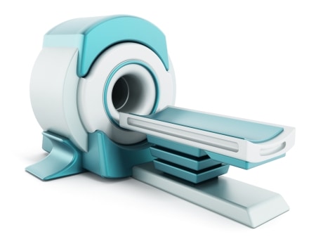
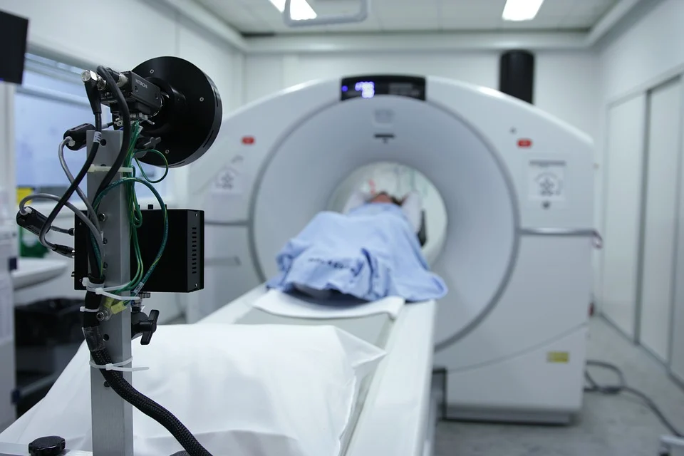
An MRI scanner is a large cylinder with powerful magnets. It has a sliding table in the centre so that a patient may be placed effortlessly into the machine. He or she remains horizontal, at rest during the entire scan procedure. Depending on the body part being scanned, the patient is moved into the scanner head or feet first. The scanner is operated by a radiographer who, after ensuring the patient is completely comfortable, controls the scanner from a few metres away. At certain times during the scan, the scanner makes loud tapping noises. This is the electric current in the scanner coils being turned on and off and is perfectly normal.
It is important to keep as still as possible during the MRI scan, to achieve the highest possible quality of the images. You might also be asked to hold your breath at times. An MRI scan of a single body part typically takes around 30 minutes.
MRI versus CT
A CT scan uses X-Rays, whilst an MRI scan uses magnets and radio waves.
An MRI scan shows tendons and ligaments, whilst a CT scan does not.
An MRI scan is preferable for examining the spinal cord.
A CT scan is better suited for cancer, pneumonia, abnormal chest x-rays and bleeding on the brain, especially after an injury.
However, a brain tumor is more clearly visible on a MRI scan.
A CT scan shows organ tears and organ injury more rapidly and thus is more suitable for trauma cases.
Broken bones and vertebrae are more clearly visible on a CT scan.
CT scans provide a better image of the lungs and organs in the chest cavity between the lungs.
How does an MRI scan work?
Much of the human body is made up of water molecules, ie hydrogen and oxygen atoms. At the centre of each hydrogen atom is a particle called a proton. Protons are like tiny magnets and sensitive to magnetic fields. When the patient lies on the table, before the procedure starts, the protons in the body line up in the same direction. Then short radio wave bursts are sent to selected body areas. When these radio waves are turned on, the protons move out of alignment. When these radio waves are turned off, the protons realign again.
The exact location of the protons in the body and the time it takes for them to move out of and back to their original alignment is detected by the MRI scanner sensors, which send out this information via radio signals. These signals, from all protons in the internal area under surveillance, combine to create an comprehensive image. The image is highly detailed and can show even the smallest abnormality.
What is a contrast agent and when is it used?
To enhance the visibility of particular tissues relevant to the scan, a contrast agent – a dye – is sometimes injected into the bloodstream. The most frequently used MRI contrast material contains gadolinium, a key component which has magnetic properties. It circulates through the blood stream and is absorbed in certain tissues, which then stand out more clearly on the scan.
Contrast material can make the patient feel warm or possibly cause a headache or dizziness. Although the dye is harmless, a patient might be asked, before it is administered, to provide eGFR blood results from within the past three months. Patients are not given contrast material if they have poor kidney function or renal impairment.
Safety of an MRI scan
Despite extensive research, no evidence has been found of any risk to the human body from the magnetic fields and radio waves that are used during MRI scans. This means that an MRI scan is one of the safest medical procedures available today. Only a claustrophobic patient might find it uncomfortable. However, the majority of patients overcome this, with support if necessary from the radiographer. Upon request, it may be possible to organise an open and/or upright scanner.
What are the risks and restrictions of an MRI Scan?
As mentioned above, an MRI scan is safe. Nevertheless, MRI scans are typically not recommended during pregnancy. Also, due to the magnetism, metallic implants or joints can affect the magnetic field and distort the images. Consequently, patients with artificial heart valves, pacemakers, metal clips, metal fragments, ear implants or insulin pumps, as well as those undergoing chemotherapy, are typically not recommended to have an MRI scan. Dependant on the body area to be scanned, guidelines may be given about eating and drinking before the scan. Where appropriate, the Imaging Centre will advise the patient about this prior to the appointment.
Types of MRI Scanners
Conventional MRI Scan
Standard 1.5 T MRI machine for regular scans
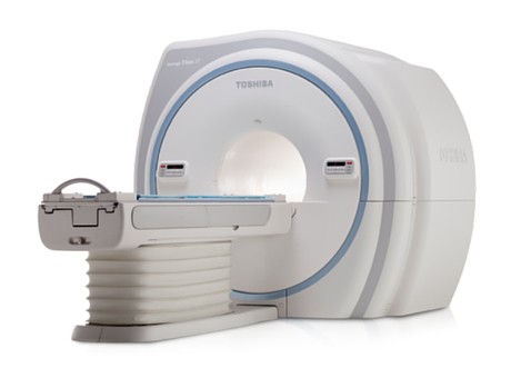
3 T MRI Scan
Higher resolution for a better imaging quality
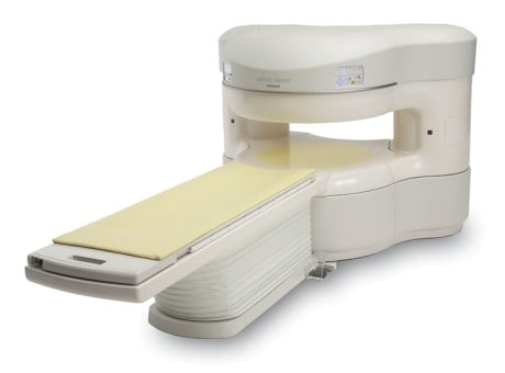
Open MRI Scan
A comfortable scan that fits any size patient
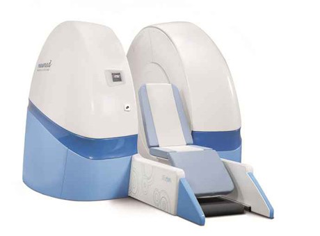
Upright MRI Scan
For claustrophobic patients and weight bearing positions

