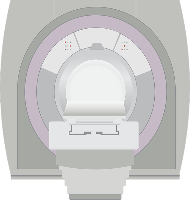
23 Feb What is the Magnetic resonance imaging (MRI)
MRI (Magnetic Resonance Imaging) scans with the use a strong magnetic field to give detailed cross-sectional images of soft tissue, muscle, nerves and blood vessels. MRI is commonly used at NHNN to diagnose many diseases and injuries including stroke and intracranial bleeds, neuro-oncology, dementia, cognitive disorders, epilepsy, movement disorders, multiple sclerosis and demyelinating diseases, CSF circulation impairment and hydrocephalus using CSF flow Phase-contrast MR techniques, neurometabolic disorders, neuromuscular disorders, infections, spinal diseases and injuries and many more.
The MRI department at NHNN also provides a unique intra-operative imaging service for patients undergoing multiple operations including DBS insertion, craniotomy and thalamotomy. The iMRI suite provides an operating theatre and MRI scanning capabilities during surgery.
Advanced neuroimaging techniques used at NHNN include fMRI (to locate specific functions in the brain), tractography (to identify white matter tracts), MR perfusion (to assess brain perfusion alterations in neurological diseases), MR spectroscopy (to assess metabolic changes in the brain) and MR neurography (for the evaluation of peripheral nerve disease), using state-of-the art post-processing tools and reporting techniques.
Information source:
www.nhs,uk
https://www.uclh.nhs.uk/our-services/find-service/neurology-and-neurosurgery/neuroradiology/magnetic-resonance-imaging-mri
[yasr_overall_rating size=”large”]


